What is amyloid?
The amyloidoses are disorders of protein folding. At least 20 different proteins can form
the extracellular deposits of non-functional β-pleated sheets that typify amyloid fibrils.
Fibrils stain with Congo red dye and produce apple-green birefringence under polarized
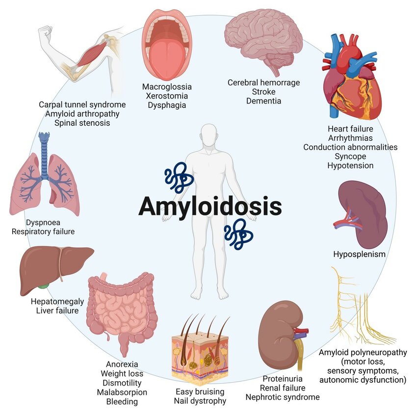
light. Amyloid protein may be deposited in any tissue, including the kidney,
gastrointestinal, heart, skin, muscle, brain, and/or blood vessels. Accumulation of protein
eventually causes organ failure (often renal).
 Reactive systemic amyloidosis (AA) develops in association with chronic inflammation,
including chronic infections, e.g. leprosy, tuberculosis, or osteomyelitis, and chronic
inflammatory diseases, e.g. RA and ankylosing spondylitis. Amyloidosis is also a
frequent complication in patients with chronic kidney disease on maintenance
haemodialysis. Rarely, systemic amyloidosis may be hereditary, e.g. as a result of
autoinflammatory syndromes or in association with FMF.
Monoclonal Ig light-chain amyloidosis (AL), which has similar clinical features to AA, is
seen in association with diseases involving B-lymphocytes such as multiple myeloma, B-
cell lymphomas, and macroglobulinaemia.
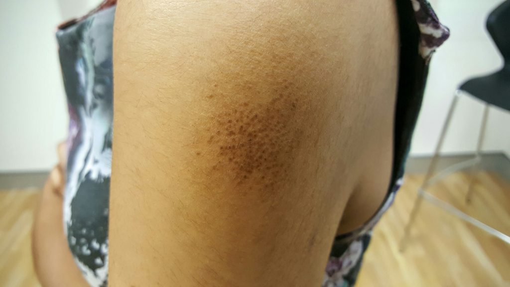
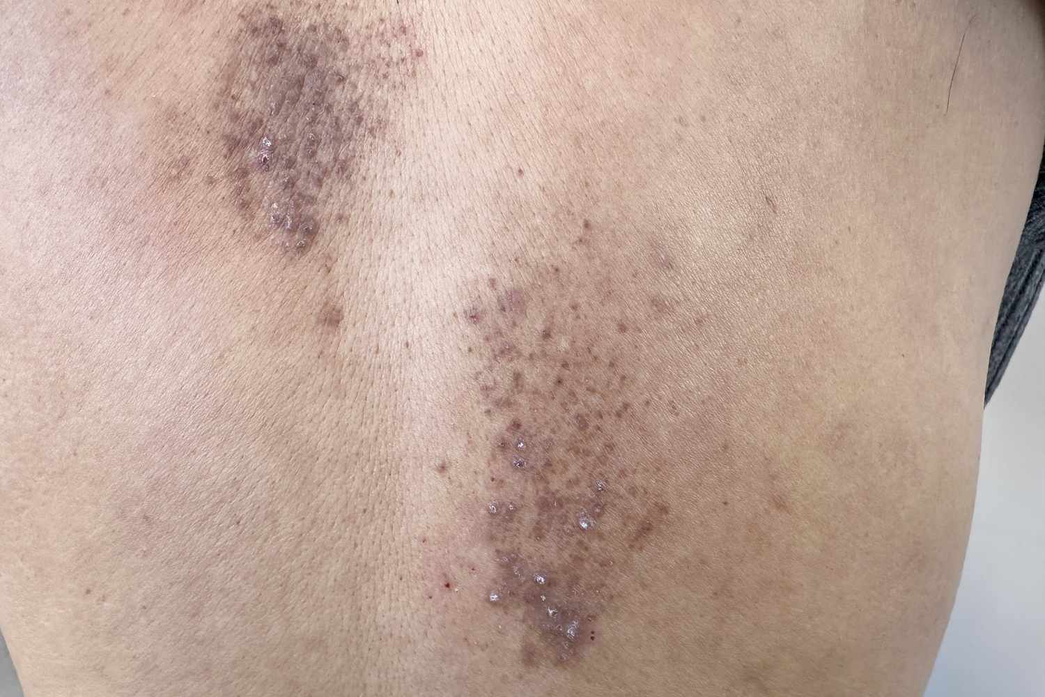
The commonest forms of primary localized cutaneous amyloidosis (macular and
lichenoid) are caused by the deposition of keratin filaments in the skin and are not
associated with systemic amyloidosis.
Systemic amyloidosis:
•Smooth yellowish or rather waxy, translucent-looking, but firm, papules, nodules, or
plaques on the face (eyelids, nasolabial folds, perioral), neck, or trunk (chest, flexures,
periumbilical). Some may be purpuric or become purpuric, if rubbed.
• Petechiae and/or purpura arising spontaneously.
• Pinch purpura: pinching or rubbing the skin causes purpura.
• Purpura of the face or neck induced by coughing or vomiting.
• Periorbital purpura, including ‘post-proctoscopy palpebral purpura’ (purpura on the
eyelids of patients who have been positioned head down for investigation by
proctoscopy).
• Mucosal infiltration:
o Smooth, pale, enlarged, indurated tongue (rare but virtually pathognomonic). the
teeth may produce scalloped indentations on the sides of the tongue.
o Thickened gingiva that bleed easily.
o Pale red or yellow papules on buccal, conjunctival, nasal, vaginal, or anal mucosa.
• Hepatosplenomegaly.
• Evidence of systemic involvement such as nephrotic syndrome, renaldisease, sensory or
autonomic neuropathy, heart failure, arrhythmias, malabsorption, or GI haemorrhage.