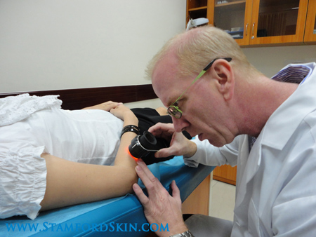

Normally, when light strikes the surface of the skin, 90% of the light is reflected off the top layers of the skin. This means that when looking at the skin by naked eye, without the use of a Dermatoscope, what the naked eye sees in a skin lesion is only the very superficial aspect of the lesion or skin. Pigmentation networks, blood vessels, and other key structures cannot be seen by the naked eye due to the reflection of light from the top layers of the skin (the ability of skin to reflect light and sun provides a protective benefit of course).
Most physicians when examining moles and other pigmented skin lesions for possible cancer or melanoma use what are called the ABCDE Clinical Criteria that can be judged by naked eye examination. A—Assymetry of the skin lesion; B—Border Irregularity of the skin lesion; C—Color Variegation in the skin lesion; D—Diameter (above 6 mm in size) of the skin lesion; E—Elevation and/or Evolution (changes noted by the patient in their moles). The use of Dermoscopy allows those physicians trained in the technique to go beyond these criteria based on naked eye examination alone. Using the Dermatoscope, when it is applied to the same skin lesions, now allows us to make out very unique and characteristic features of a pigmented skin lesion and even non-pigmented skin lesions. With this technique, features characteristic of a Melanoma (a worrisome Skin Cancer), features characteristic of Basal Cell Skin Cancer, Squamous Cell Cancer, and features of many other suspicious and worrisome skin lesions can be appreciated sooner in their evolution, at an earlier time and when it comes to Skin Cancer Detection, especially with Melanoma, the earlier the lesion is identified and removed the better the prognosis.
This may not even be the best aspect of the use of Dermoscopy, however. There are several benign or normal skin lesions that by naked eye examination alone may share some challenging characteristics or features (in the ABCDE guideline) that suggest they could possibly be a Skin Cancer. Using the Dermatoscope on these often squarely identifies the lesion as definitely benign. Both benign and atypical moles, and other pigmented but benign lesions like irregular freckles, unusual sun spots, odd-looking seborrhoeic keratoses, and hemanigiomas can be definitely distinguished as benign with the Dermatoscope.
It has been shown that the number of benign skin lesions removed by a Dermatologist who does not have the use of a Dermatoscope is 40 benign lesions to every one malignant lesion that is found by naked eye examination. That represents a lot of benign skin lesions that need removal to find the one cancerous lesion. When Dermoscopy is employed by a physician who is trained in the use of the Dermatoscope and pattern-recognition of various benign and malignant lesions this ratio then drops down to 4 benign lesions removed for every one malignant skin cancer removed. Where does the difference come in? It is explained by the ability to identify those lesions that by naked eye exam that appear worrisome and would otherwise be removed or biopsied, that when examined under the Dermatoscope can be truly identified as having benign features and thus do not need to be removed, meaning less biopsies, and less minor skin surgery for patients that are followed by physicians employing Dermoscopy.